 Talk to Sales
 Benchmarks
View scores and output across OCR models spanning many document categories.
Want to run these evals on your own documents?
Talk to Sales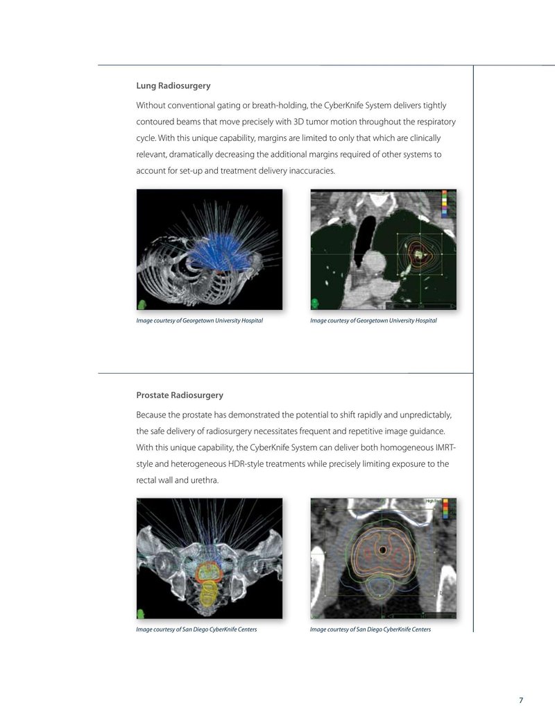
Lung Radiosurgery
Without conventional gating or breath-holding, the CyberKnife System delivers tightly contoured beams that move precisely with 3D tumor motion throughout the respiratory cycle. With this unique capability, margins are limited to only that which are clinically relevant, dramatically decreasing the additional margins required of other systems to account for set-up and treatment delivery inaccuracies.

Image courtesy of Georgetown University Hospital

Image courtesy of Georgetown University Hospital
Prostate Radiosurgery
Because the prostate has demonstrated the potential to shift rapidly and unpredictably, the safe delivery of radiosurgery necessitates frequent and repetitive image guidance. With this unique capability, the CyberKnife System can deliver both homogeneous IMRT-style and heterogeneous HDR-style treatments while precisely limiting exposure to the rectal wall and urethra.

Image courtesy of San Diego CyberKnife Centers

Image courtesy of San Diego CyberKnife Centers
7