 Talk to Sales
 Benchmarks
View scores and output across OCR models spanning many document categories.
Want to run these evals on your own documents?
Talk to Sales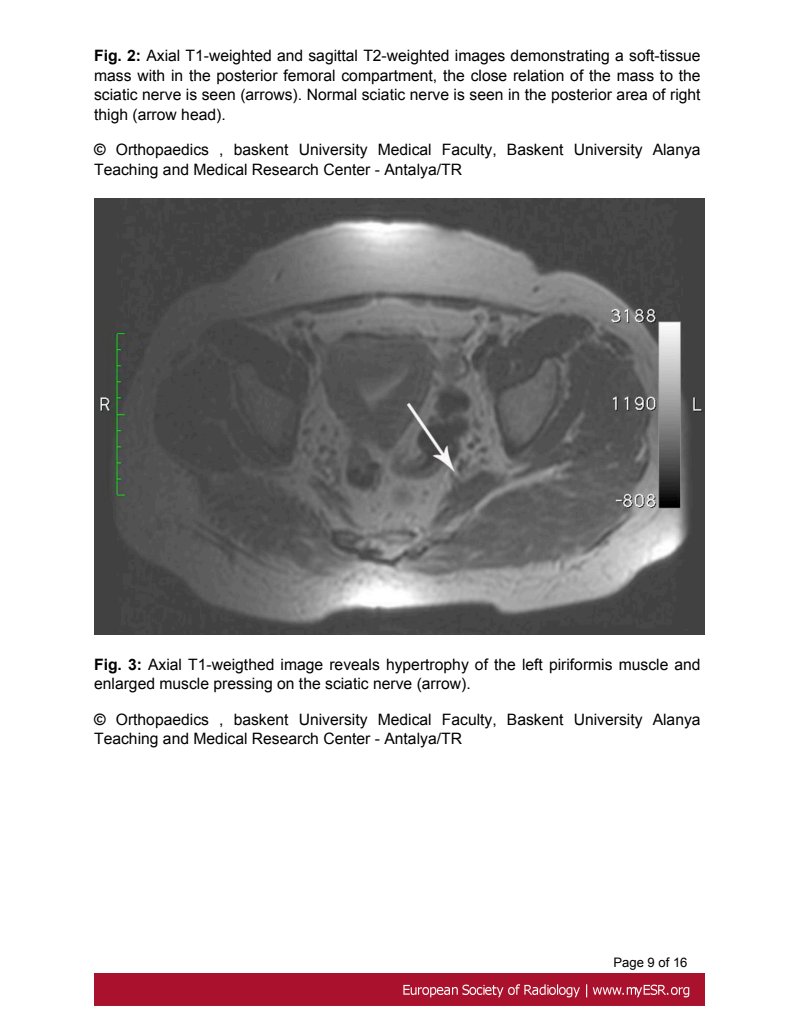
Fig. 2: Axial T1-weighted and sagittal T2-weighted images demonstrating a soft-tissue mass within the posterior femoral compartment, the close relation of the mass to the sciatic nerve is seen (arrows). Normal sciatic nerve is seen in the posterior area of right thigh (arrow head).
© Orthopaedics , baskent University Medical Faculty, Baskent University Alanya Teaching and Medical Research Center - Antalya/TR

Fig. 3: Axial T1-weighted image reveals hypertrophy of the left piriformis muscle and enlarged muscle pressing on the sciatic nerve (arrow).
© Orthopaedics , baskent University Medical Faculty, Baskent University Alanya Teaching and Medical Research Center - Antalya/TR
Page 9 of 16
European Society of Radiology | www.myESR.org