 Talk to Sales
 Benchmarks
View scores and output across OCR models spanning many document categories.
Want to run these evals on your own documents?
Talk to Sales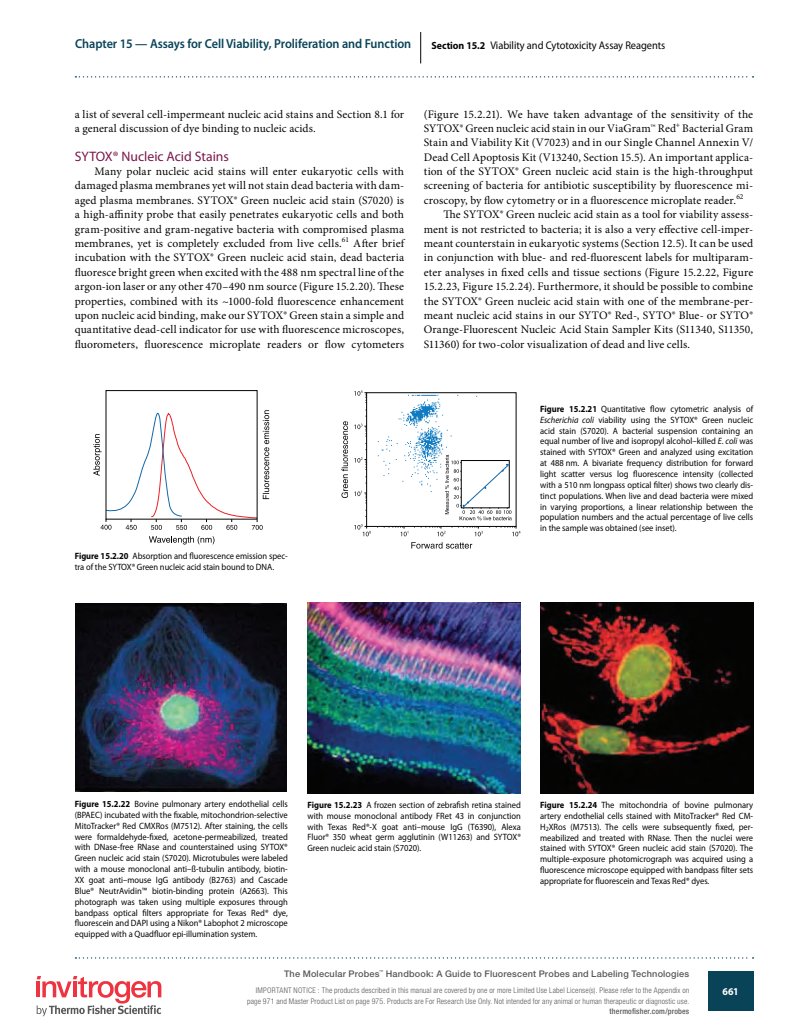
Chapter 15 — Assays for Cell Viability, Proliferation and Function
Section 15.2 Viability and Cytotoxicity Assay Reagents
a list of several cell-impermeant nucleic acid stains and Section 8.1 for a general discussion of dye binding to nucleic acids.
SYTOX® Nucleic Acid Stains
Many polar nucleic acid stains will enter eukaryotic cells with damaged plasma membranes yet will not stain dead bacteria with damaged plasma membranes. SYTOX® Green nucleic acid stain (S7020) is a high-affinity probe that easily penetrates eukaryotic cells and both gram-positive and gram-negative bacteria with compromised plasma membranes, yet is completely excluded from live cells. 61 After brief incubation with the SYTOX® Green nucleic acid stain, dead bacteria fluoresce bright green when excited with the 488 nm spectral line of the argon-ion laser or any other 470–490 nm source (Figure 15.2.20). These properties, combined with its ~1000-fold fluorescence enhancement upon nucleic acid binding, make our SYTOX® Green stain a simple and quantitative dead-cell indicator for use with fluorescence microscopes, fluorometers, fluorescence microplate readers or flow cytometers
(Figure 15.2.21). We have taken advantage of the sensitivity of the SYTOX® Green nucleic acid stain in our ViaGram™ Red Bacterial Gram Stain and Viability Kit (V7023) and in our Single Channel Annexin V/ Dead Cell Apoptosis Kit (V13240, Section 15.5). An important application of the SYTOX® Green nucleic acid stain is the high-throughput screening of bacteria for antibiotic susceptibility by fluorescence microscopy, by flow cytometry or in a fluorescence microplate reader. 62
The SYTOX® Green nucleic acid stain as a tool for viability assessment is not restricted to bacteria; it is also a very effective cell-impermeant counterstain in eukaryotic systems (Section 12.5). It can be used in conjunction with blue- and red-fluorescent labels for multiparameter analyses in fixed cells and tissue sections (Figure 15.2.22, Figure 15.2.23, Figure 15.2.24). Furthermore, it should be possible to combine the SYTOX® Green nucleic acid stain with one of the membrane-permeant nucleic acid stains in our SYTO® Red-, SYTO® Blue- or SYTO® Orange-Fluorescent Nucleic Acid Stain Sampler Kits (S11340, S11350, S11360) for two-color visualization of dead and live cells.

Figure 15.2.20 Absorption and fluorescence emission spectra of the SYTOX® Green nucleic acid stain bound to DNA.

Figure 15.2.21 Quantitative flow cytometric analysis of Escherichia coli viability using the SYTOX® Green nucleic acid stain (S7020). A bacterial suspension containing an equal number of live and isopropyl alcohol-killed E. coli was stained with SYTOX® Green and analyzed using excitation at 488 nm. A bivariate frequency distribution for forward light scatter versus log fluorescence intensity (collected with a 510 nm longpass optical filter) shows two clearly distinct populations. When live and dead bacteria were mixed in varying proportions, a linear relationship between the population numbers and the actual percentage of live cells in the sample was obtained (see inset).

Figure 15.2.22 Bovine pulmonary artery endothelial cells (BPAEC) incubated with the fixable, mitochondrion-selective MitoTracker® Red CMXRos (M7512). After staining, the cells were formaldehyde-fixed, acetone-permeabilized, treated with DNase-free RNase and counterstained using SYTOX® Green nucleic acid stain (S7020). Microtubules were labeled with a mouse monoclonal anti- -tubulin antibody, biotin-XX goat anti-mouse IgG antibody (B2763) and Cascade Blue® NeutrAvidin™ biotin-binding protein (A2663). This photograph was taken using multiple exposures through bandpass optical filters appropriate for Texas Red® dye, fluorescein and DAPI using a Nikon® Labophot 2 microscope equipped with a Quadfluor epi-illumination system.

Figure 15.2.23 A frozen section of zebrafish retina stained with mouse monoclonal antibody FRet 43 in conjunction with Texas Red®-X goat anti-mouse IgG (T6390), Alexa Fluor® 350 wheat germ agglutinin (W11263) and SYTOX® Green nucleic acid stain (S7020).

Figure 15.2.24 The mitochondria of bovine pulmonary artery endothelial cells stained with MitoTracker® Red CM-H XRos (M7513). The cells were subsequently fixed, permeabilized and treated with RNase. Then the nuclei were stained with SYTOX® Green nucleic acid stain (S7020). The multiple-exposure photomicrograph was acquired using a fluorescence microscope equipped with bandpass filter sets appropriate for fluorescein and Texas Red® dyes.
invitrogen
by Thermo Fisher Scientific
The Molecular Probes™ Handbook: A Guide to Fluorescent Probes and Labeling Technologies
IMPORTANT NOTICE: The products described in this manual are covered by one or more Limited Use Label License(s). Please refer to the Appendix on page 971 and Master Product List on page 975. Products are For Research Use Only. Not intended for any animal or human therapeutic or diagnostic use.
thermofisher.com/probes
661